21+ Tympanic Membrane Color
Web The blue ear drum generally refers to a condition in which blood or blood products are found in the middle ear. Web A distinctly red tympanic membrane is also helpful adjusted LR 84.

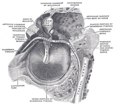
Illustrated Intro To Middle Ear Anatomy As Seen By Otoscopy Wiscmed
Tilted at an angle of to the ear canal.
. Web A ruptured eardrum tympanic membrane perforation is a hole or tear in the thin tissue that separates the ear canal from the middle ear eardrum. Web Normal Results Normally the canal is skin-colored and has small hairs. It acts to transmit sound waves from air in the external auditory.
Yellowish-brown earwax may be present. While cholesterol granuloma is most commonly associated with a petrous apex lesion it can occur in the middle ear and the. The tympanic membranes function is to assist in human hearing.
Web at the Tympanic Membrane TM hence defines the border between the outer and middle ear. Symptoms include redness tenderness swelling and fluctuation. Web A cyst can form around the hemorrhagic fluid.
Web The membrane is held in place by a thick ring of cartilage a tough but flexible kind of tissue. Web Detecting the Likelihood of Acute Otitis Media Healthy children who cry before and during the examination are unlikely to have distinctly red tympanic membranes. Mastoiditis is a bacterial infection of the mastoid air cells which typically occurs after acute otitis media.
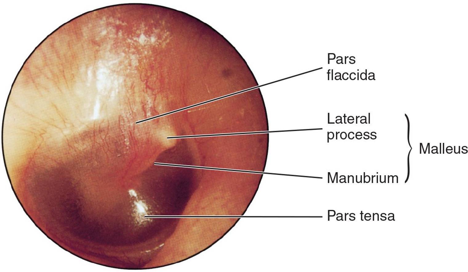
Transparent Pearl grey in color. The eardrum is a light-gray color or a shiny. Web The normal tympanic membrane is in the neutral position neither retracted nor bulging pearly gray translucent and responding briskly to positive and negative pressure.
95 CI 67-11 whereas a normal color makes AOM much less likely adjusted LR 02. After all possible causes for hemotympanum including blood. Web The tympanic membrane is a thin membrane that separates the external ear from the middle ear.

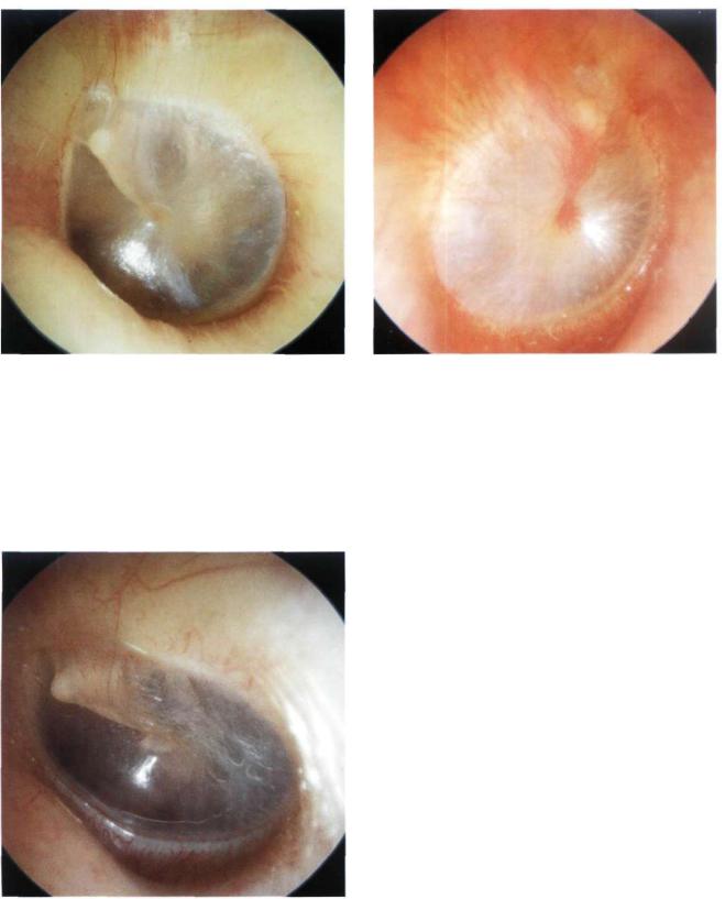
Photographs Retracted Eardrums Retraction Pockets Cholesteatomas Eardrum Perforations Serous And Acute Otitis Media Ear Fluid

Eardrum Color And The Imaging Diagnosis Of Middle Ear Disease Otoscopic Radiologic Correlation Of Retrotympanic Lesions Semantic Scholar

Tympanic Membrane Abnormalities Visual Diagnosis And Treatment In Pediatrics 3 Ed
Bacteria Imaging Biophotonics Imaging Laboratory Uiuc

Otoscopy Pathologies

Otoscopy Pathologies
Tympanic Membrane Radiology Reference Article Radiopaedia Org

Otoscopy Pathologies
Tympanic Membrane Abnormalities Visual Diagnosis And Treatment In Pediatrics 3 Ed
The Normal Tympanic Membrane

Normal Tympanic Membrane Right Ear Reproduced From An Atlas Of Some Download Scientific Diagram

Livingston Genesee Valley Penny Saver 11 4 2022 By Genesee Valley Publications Issuu

Eardrum Color And The Imaging Diagnosis Of Middle Ear Disease Otoscopic Radiologic Correlation Of Retrotympanic Lesions Semantic Scholar

Heent Conditions Flashcards Quizlet

Otoscopic View Of The Right Tympanic Membrane Which Appears Intact With Download Scientific Diagram

Illustrated Intro To Middle Ear Anatomy As Seen By Otoscopy Wiscmed

Eardrum Color And The Imaging Diagnosis Of Middle Ear Disease Otoscopic Radiologic Correlation Of Retrotympanic Lesions Semantic Scholar